The Shroud of Turin · Chapter 12 of 21
The crucifixion: the wounds of the hands, feet, and back
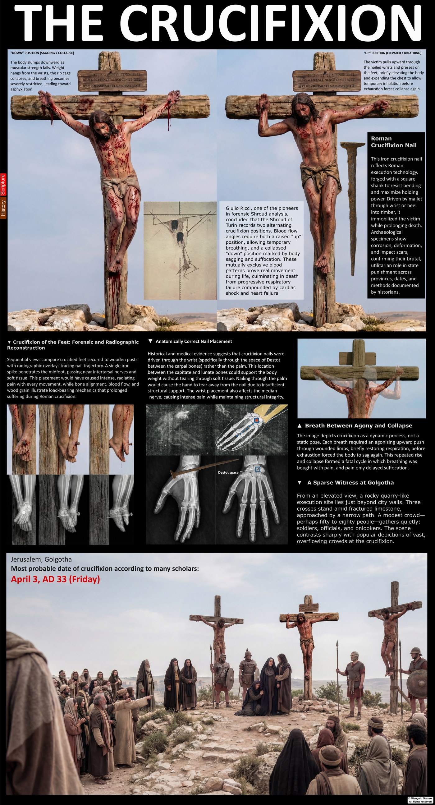
The Gospels describe the crucifixion in a few plain sentences. They name the place, the soldiers, the inscription, and the two men crucified with Jesus, and they record his last words. They say little about the nails, the position of the body, or the wounds themselves. The cloth records the wounds in a form that medical and forensic writers have studied in detail, and this chapter examines the readings of the hands, the feet, and the back, and what each can and cannot show.
What the Gospels say
The Gospels report that the soldiers stripped Jesus and divided his garments, casting lots for the tunic (Matthew 27:35; Mark 15:24; Luke 23:34; John 19:23–24). John says that the tunic was seamless and that the soldiers did not tear it (John 19:23). The cross was set up, and an inscription was placed above him (Mark 15:26; John 19:19). The Gospels do not mention the nails in the body or the order in which they were driven, and John records that the soldiers did not break his legs, which was the usual practice for hastening death (John 19:31–33).1
The Gospel accounts do not describe the cross in detail, and the reader depends on other sources for its form. Roman crucifixion varied in practice, and the surviving evidence is limited. The only Roman-era crucifixion victim known from archaeology, Jehohanan, was found in a Jerusalem ossuary in 1968, with a nail through the heel bones.2
Roman practice
The condemned man usually carried the horizontal beam, the patibulum, to the execution site. The upright stake was normally fixed there. The victim was then fixed to the cross, most often with nails, and sometimes with ropes. The Roman sources describe the punishment as slow and public, and they say that death could take hours or days.3
The wrists and the nails
Medieval and Renaissance art almost always shows nails through the palms. The cloth, on the other hand, shows the bloodstains on the wrists, not the palms.4 Pierre Barbet tested the question in the 1930s with cadaver experiments. He found that a nail through the palm tears under the weight of a suspended body, and he argued that the wrist, through the carpal space called Destot’s space, is a more stable point of support.4
The wrist placement has a consequence for the nerves. A nail in the wrist can injure the median nerve, which supplies the muscles of the thumb. Barbet proposed that such an injury causes the thumb to draw into the palm, and that this explains the absence of visible thumbs on the cloth. Other writers have pointed out that a median nerve injury usually causes paralysis of the thumb muscles rather than contraction, and that the natural resting position of a hand in death can also hide the thumb.5 The cause is debated. The fact that the thumbs are not visible on the cloth is not in dispute.
The feet
Medieval depictions usually show the feet nailed separately, side by side. The cloth shows the feet overlapped, one over the other. Rigor mortis, the stiffening after death, would tend to keep the limbs in the position they held when the body was taken down. On that reading, the overlapped feet point to a single nail through both feet.4
The path of such a nail is debated. Barbet proposed a route between the long bones of the foot. More recent models suggest a path through the tarsal bones of one foot and the metatarsals of the other. A bloodstain near the right heel has been read as a second, heel wound. The stain is diffuse, and it can also be explained by blood that pooled and drained after death, spreading into the linen by capillary action. The heel question is open.5
The back and the scourge marks
The dorsal image shows the dumbbell-shaped marks of the scourging described in Chapter 8. The number of marks has been counted in several ways. Earlier counts gave roughly 90 to 120 marks on the back, and later analyses of the same photographs report several hundred, as Chapter 8 explains. The disagreement is about counting and about which marks are accepted, not about whether the marks are there.6
The scourging is also relevant to the crucifixion. A flogging of this severity causes blood loss and shock, and a man who has been flogged so severely is already weakened when he reaches the cross. The physical state of the victim on the cross therefore depended on what came before it.
The blood flows on the arms
The bloodstains on the forearms run in more than one direction. Some are steep and nearly vertical, and others are more horizontal. Zugibe and others have read this as evidence that the body changed position during the crucifixion, hanging when slumped and lifting when the victim pushed up on the nailed feet. On this reading the blood follows the changing angle of the arms.5 The reading is interpretive, and it depends on the assumed position of the body at the time the blood ran. Other factors, including the way the cloth was laid over the arms, also affect the flow.
Breathing on the cross
The question of breathing is central to the mechanism of death, and it is discussed in Chapter 13. The positional blood flows are often used in that discussion. Zugibe’s experiments with suspended volunteers found that breathing shifted toward the abdomen, that the chest became relatively fixed, and that oxygen levels did not fall in the short trials. He later argued that shock and trauma may have played a larger role than asphyxia. The positional reading of the blood does not decide that question by itself.5
The argument from the wounds
Several features of the cloth diverge from the artistic tradition: the wrist placement, the absent thumbs, the overlapped feet, and the blood flows that change with position. Each has been read as consistent with a real crucifixion. Together they form a coherent picture, and the picture is hard to reproduce by artistic convention.
The argument has limits. A forger who had seen relics or real wounds could have copied some of these features, and a forger who had studied the anatomy could have chosen wrist nailing. The forensic reading shows consistency with a crucifixion; it does not prove when the cloth was made. The 1988 radiocarbon result places the linen in the medieval period, and that result is contested, as discussed in Chapter 19.7
What the wounds can and cannot show
The wounds can show that the hands and feet were pierced in a way consistent with fixing a body to a cross, that the back bore a dense pattern of scourge marks, and that the blood flows on the arms record changes in position. The readings of the wrists, the thumbs, the feet, and the flows are informed by medical and forensic analysis, and the chapter has set out where each is firm and where it is contested.
They cannot tell us the length of the crucifixion, the number of nails, the exact form of the cross, or the reasons for the particular placements. Those questions are addressed, as far as the evidence allows, in the chapters on the cause of death and the burial.
References
- Matthew 27:35; Mark 15:24, 26; Luke 23:34; John 19:19, 23–24, 31–33. John 19:23–24; John 19:31–33.
- Jehohanan ben Hagkol, Jerusalem ossuary, discovered 1968. Publication details to be confirmed before publication.
- Roman crucifixion practice as discussed in the medical and historical literature. See Edwards, W. D., Gabel, W. J., & Hosmer, F. E. (1986). On the physical death of Jesus Christ. JAMA, 255(11), 1455–1463. doi:10.1001/jama.1986.03370110077025
- Barbet, P. (1953). A Doctor at Calvary. New York: P. J. Kenedy & Sons.
- Zugibe, F. T. (2005). The Crucifixion of Jesus: A Forensic Inquiry. New York: M. Evans and Company.
- Scourge-mark counts as reported in the forensic literature on the Shroud. See Chapter 8 for the sources and the range of counts.
- Damon, P. E., et al. (1989). Radiocarbon dating of the Shroud of Turin. Nature, 337, 611–615. doi:10.1038/337611a0