The Shroud of Turin · Chapter 1 of 21
What Is the Shroud of Turin?
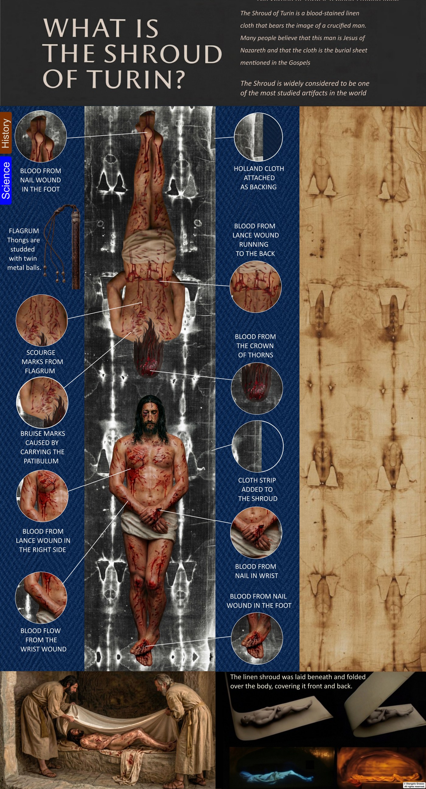
It is fourteen feet long. A man’s form lies across it, front and back, head to head, as if the linen had been draped over a body and had settled into every contour of it.
The Shroud of Turin is a rectangular length of linen, about 4.36 metres (14 feet 3 inches) long and 1.10 metres (3 feet 7 inches) wide. It is kept in the Cathedral of Saint John the Baptist in Turin, Italy, and it is one of the most intensively studied textiles in the world. It carries a faint image of a crucified man and reddish-brown stains that many observers identify as blood. Many Christians believe it is the burial cloth of Jesus of Nazareth, the linen the Gospels describe as wrapped around his body after his death.
This chapter sets out what the cloth is, what has been measured on it, how its study developed, and which questions any serious explanation has to answer. Everything after it builds on the material described here.
The cloth
The linen is the colour of old tea. It is woven in a herringbone twill, a three-to-one pattern in which each weft thread passes over three warp threads before it goes under one. The weave catches the light differently depending on how the cloth is held, and it is the first thing a textile examiner notices. The fibres are flax. Whether the weave points to the first century or to the medieval period is one of the textile questions specialists have argued over for decades. Chapter 2 takes it up.
The cloth has a documented physical history. The most visible event is the fire of 1532, when the silver reliquary in which it was kept, at the Sainte-Chapelle in Chambéry, caught fire.1 Molten silver fell onto the folded linen, and the water used to put out the flames soaked parts of it. The result is a set of scorch lines and water stains on both the front and back, and a series of patches sewn on by the nuns who repaired the damage. The pattern of scorches and patches matches the way the cloth had been folded when the fire broke out, so the marks also record how it was stored.
In 1534 the Poor Clares of Chambéry sewed a backing of Holland linen to the cloth. Any study of the back image has to account for that backing. Later restoration work, and the fire in the Guarini Chapel in Turin in 1997, where the cloth was then kept, are further parts of its documented history.
The documented custody follows clear stages. The cloth first appears in the historical record at Lirey, in north-central France, in the 1350s, when it was shown by the Charny family. In 1389 a bishop of Troyes wrote a memorandum claiming that an artist had confessed to painting it, a claim that later scholarship has examined closely.2 The cloth passed to the House of Savoy in 1453, was taken to Turin in 1578, and remained in the Savoy family until 1983, when it was bequeathed to the Holy See.1 Its custody today rests with the Archbishop of Turin, and it is shown only on rare occasions.
The image
From about three metres away the image coheres: a bearded face, arms crossed at the groin, the long lines of the torso, and the shadows of ribs and muscle. Step closer and it breaks apart into individual threads. Each thread is discoloured only on its uppermost surface, and the colour does not soak into the fibre. What looks continuous from a distance is, up close, a pattern made of surfaces.
Four properties of the image have made it the centre of scientific debate.
1. It is not paint
No paint, dye, or pigment of any known kind has been identified in the image. There are no brush marks, no binding medium, and no layer of colour sitting on the fibres. The colour comes from a superficial change in the linen itself, confined to the outermost fibrils, which are layers only fractions of a micrometre thick. The inner fibres of each thread are essentially untouched. The 1978 scientific examination of the cloth concluded that the image results from oxidative degradation of the linen cellulose.3 Walter McCrone argued instead that the colour came from iron-oxide pigment, a view that later STURP and independent work did not support.4 The question of what produced the colour remains one of the central problems of the subject.
2. It is a negative
The image is a photographic negative: the lightest parts of the body appear dark and the darkest appear light. This was not known until 1898, when the Italian photographer Secondo Pia exposed the first photographs of the cloth and saw on the negative plate a lifelike positive portrait.5 Chapter 5 tells that story.
3. It encodes distance
When image density is read as a measure of distance, a coherent three-dimensional relief of a human body appears. Darker tones correspond to areas where the cloth lay closer to the body, and lighter tones to areas where it lay farther away. In the 1970s the American researchers John Jackson and Eric Jumper used a VP-8 image analyser, a device developed for processing spacecraft imagery, to convert the image into a three-dimensional map. Their 1984 paper is among the most cited studies of the cloth’s relief.6 How a flat surface could carry this information is one of the open problems of the subject, and Chapter 6 examines it.
4. It is superficial
Because the discolouration is confined to the surface of the fibrils, it does not behave like a stain. Microscopy and chemical tests have found that the image does not penetrate the thread the way paint or dye would. It is shallow, even, and resistant to solvents that would dissolve a pigment. That surface character is why the image is often compared to a photographic effect rather than to any ordinary artistic medium.
The stains
Across the image lie reddish-brown stains concentrated on the scalp, the wrists, the feet, the side of the chest, the shoulders, and the back. They show flow patterns, clot rings, and serum separation, which are features of liquid blood that has run from wounds and dried. Chapter 3 is devoted to them. Two findings are central here.
The first is sequence. Chemical examination reported in the scientific literature found that the blood was deposited before the body image formed. The faint body image is absent beneath the bloodstains, so the blood was on the cloth first and the image formed afterward.7
The second is serum. Under ultraviolet light several bloodstains show pale rings around their edges. When blood clots, the clot contracts and pushes clear serum outward, and the rings are the traces of that process. Such halos are difficult to reproduce artificially, which is one reason researchers regard the stains as genuine blood rather than applied pigment.
Early immunochemical work by Baima Bollone and colleagues in 1981 reported human haemoglobin and blood-group markers in the stains.8 Later work reported a blood group of AB, a result that has been examined and debated since.
The wounds that the stains describe are the basis of the cloth’s place in forensic discussion. Each is treated in its own chapter in this series:
- The scalp. The stains form a cap-shaped pattern from the forehead over the crown and down the nape, rather than a ring of punctures. Their flow patterns are compared with blood on a living scalp.
- The hands. Both hands are crossed and the thumbs are not visible. Forensic physicians have linked this to injury to the median nerve, a feature that a medieval artist working from imagination would not be likely to reproduce.9
- The wrists. The wounds lie on the wrists rather than in the palms. Early medical writing on crucifixion argued that a nail through the wrist could carry the weight of a body, whereas a nail through the palm would tear.10
- The feet. The feet carry wounds whose arrangement is discussed in relation to how they were fixed to the upright.
- The side. A stain on the right side of the chest has been read as the trace of a wound from a lance. On the back image, the separation of blood and serum is compared with the Gospel account of blood and water flowing from the side of Jesus (John 19:34).
- The back. The shoulders, back, and legs show marks that are interpreted as scourging, which is compared with the Gospel description of the flagellation.
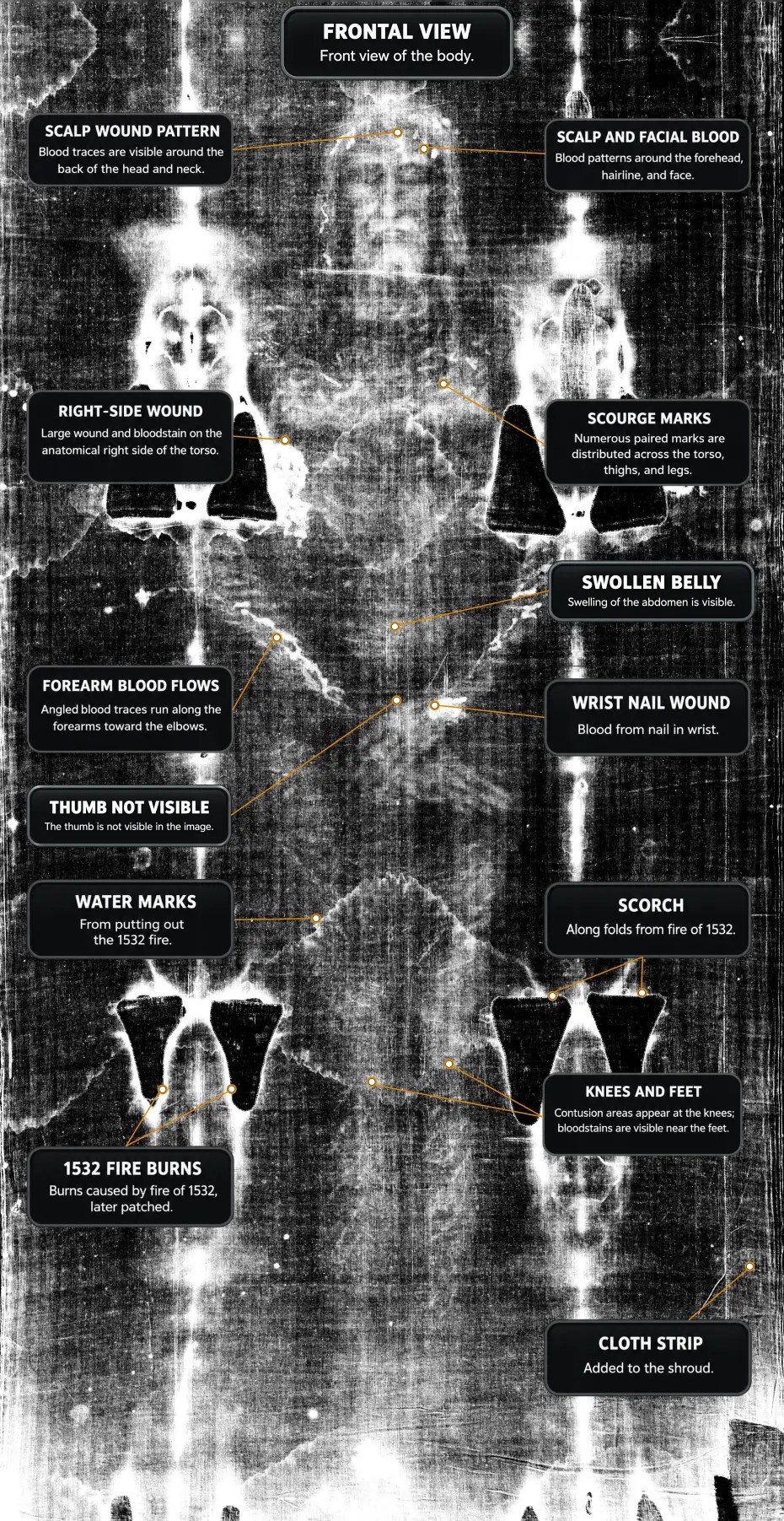
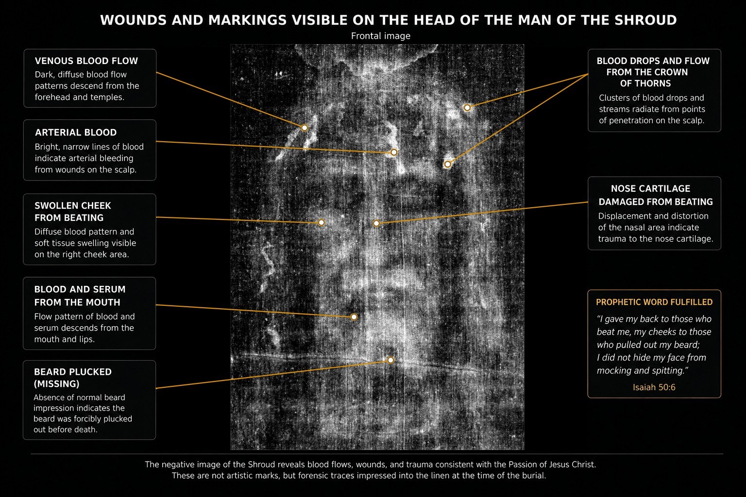
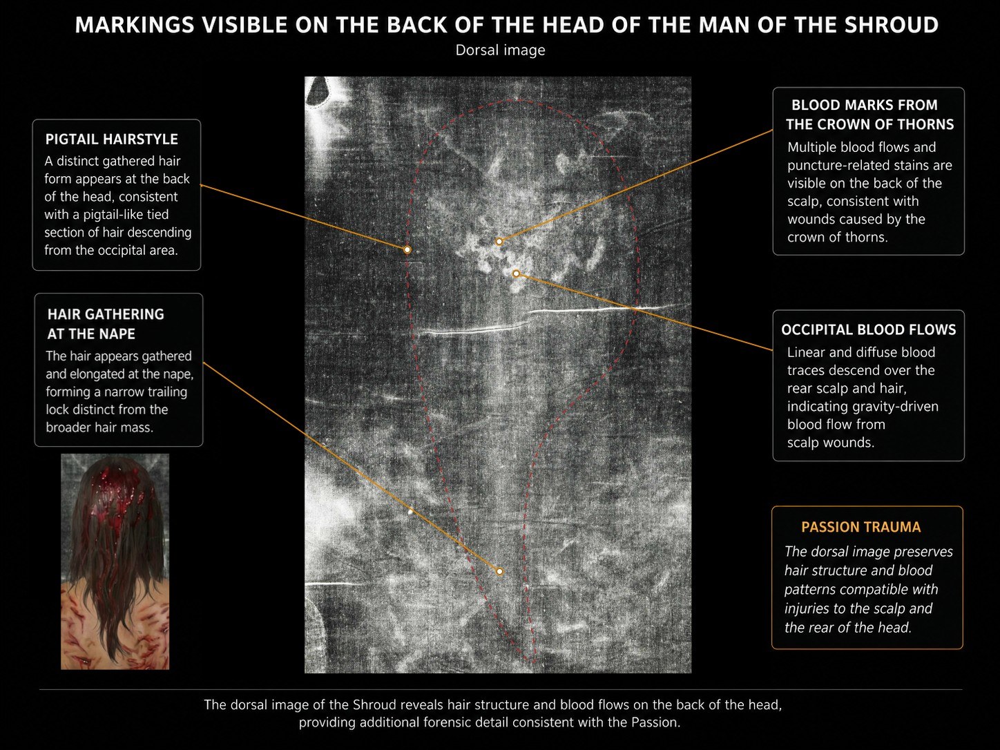
These are interpretations of the wounds, not direct measurements. The chapters on the scourging, the crown of thorns, and the crucifixion set out the evidence and the arguments against each reading.
The first scientific examinations
Modern study of the cloth began in 1898, when Secondo Pia photographed it during a public exhibition in Turin.5 His negative revealed the positive portrait and turned the body image into a matter of public attention. In 1931 Giuseppe Enrie made a second set of photographs that confirmed the negative property and allowed closer study of the anatomy.11
The next major event was the 1978 Shroud of Turin Research Project (STURP). A team of American scientists, with colleagues from Italy and elsewhere, spent about 120 hours examining the cloth in October 1978. They used spectroscopy, microscopy, X-ray fluorescence, ultraviolet, infrared, and multi-spectral photography, along with tape sampling of the fibres. The principal published summary, by Schwalbe and Rogers in 1982, is cited above.3 The team did not agree on every point, and its findings have been debated since. Chapter 18 covers the investigation.
In 1988 three laboratories in Arizona, Oxford, and Zurich dated a sample taken from one corner of the cloth by accelerator mass spectrometry. Their combined result, published in Nature in 1989, gave a calibrated calendar age of AD 1260–1390.12 That result has defined public debate about the cloth ever since, and any explanation of the Shroud has to deal with it. Chapter 19 examines the result, the objections raised against it, and the responses to those objections.
The radiocarbon date, the STURP findings, the properties of the image, and the wounds are different kinds of evidence about the same object. This series asks how an explanation can account for all of them at once.
How this series examines the evidence
The series rests on a few working principles. Primary physical data takes precedence: the chemistry of the blood, its relation to the image, the anatomical coherence of the wounds, and the sequence of clotting and flow. Environmental traces such as pollen and mineral residues are treated as corroboration or constraint rather than independent proof. Historical and iconographic material is treated as context, useful where it agrees with the physical findings and not decisive where it does not.
A hypothesis is judged by whether it accounts for all the known constraints at once. An explanation that fits one kind of evidence while contradicting another is treated as incomplete. Explanations that require knowledge or techniques unavailable at the time of the cloth’s presumed origin are examined with particular care, because anachronism is one of the most reliable signs of error.
The language is meant to be restrained. Consistency is not proof, improbability is not impossibility, and the best-fitting explanation is not an established fact. Where the evidence is silent, that silence is stated. The mechanism of image formation has no complete explanation, and it is presented here as a set of constraints and open questions.
The series also states its own position. This site is run by a Christian believer, and the faith dimension is kept separate from the forensic chapters. The closing chapters address what the evidence might mean for faith, and they are offered as an invitation rather than a conclusion drawn from the physical data.
Three questions
Three questions organise the rest of the series:
- Who was this man? The wounds, stains, posture, and burial traces are read as the record of a specific execution. The chapters on the Passion follow that record from Gethsemane to the tomb.
- How did his image form? The image is superficial, non-pigmented, a negative, and it encodes distance. Any explanation has to account for each property, and the chapters on image formation test the candidate explanations against them.
- When did this cloth exist? The radiocarbon result places the cloth in the medieval period. The documentary history, the textile evidence, the pollen and mineral traces, and the image itself are read by some researchers as pointing elsewhere. This is the most contested of the three questions.
None of these questions has a settled answer, and none can be dismissed. The series treats each as open and follows the evidence layer by layer, discipline by discipline.
The first layer is the linen itself. Where was it made, and when? That is the subject of Chapter 2.
References
- Wilson, I. (1978). The Turin Shroud. London: Victor Gollancz.
- d’Arcis, P. (1389). Memorandum to Clement VII concerning the Lirey cloth. Cited in later historical studies of the Shroud’s early exhibition.
- Schwalbe, L. A., & Rogers, R. N. (1982). Physics and chemistry of the Shroud of Turin: A summary of the 1978 investigation. Analytica Chimica Acta, 135, 3–49.
- McCrone, W. C. (1990). The Shroud of Turin: Blood or artist’s pigment? Accounts of Chemical Research, 23(3), 77–83.
- Pia, S. (1898). The first photograph of the Holy Shroud.
- Jackson, J. P., Jumper, E. J., & Ercoline, W. R. (1984). Correlation of image intensity on the Turin Shroud with the 3-D structure of a human body shape. Applied Optics, 23(14), 2244–2270.
- Heller, J. H., & Adler, A. D. (1981). A chemical investigation of the Shroud of Turin. Canadian Society of Forensic Science Journal, 14(3), 81–103.
- Baima Bollone, P., Jorio, M., & Massaro, A. L. (1981). La dimostrazione della presenza di tracce di sangue umano sulla Sindone. Sindon, 30, 5–8.
- Zugibe, F. T. (2005). The Crucifixion of Jesus: A Forensic Inquiry. New York: M. Evans.
- Barbet, P. (1953). A Doctor at Calvary. New York: Image Books.
- Enrie, G. (1931). Photographic survey of the Holy Shroud, Turin.
- Damon, P. E., et al. (1989). Radiocarbon dating of the Shroud of Turin. Nature, 337, 611–615.
Further resources
- shroud.com, the archive built by Barrie Schwortz, the STURP documentarian, with the 1978 archive, technical papers, and a photographic library.
- sindone.org, the International Center of Sindonology in Turin, with academic resources and official updates from the custodians of the cloth.
- museosindone.it, the official museum site, with material on the history and conservation of the cloth.
- sturp.org, the archive of the 1978 Shroud of Turin Research Project.
- shroudphotos.com, Vernon Miller’s STURP photographs, the primary photographic reference for much of the research.
- avvolti.org, an interactive multimedia guide to the image, layer by layer.
- sindonology.org, an independent platform with translations and symposium reports.