The Shroud of Turin · Chapter 3 of 21
The blood of the Shroud of Turin
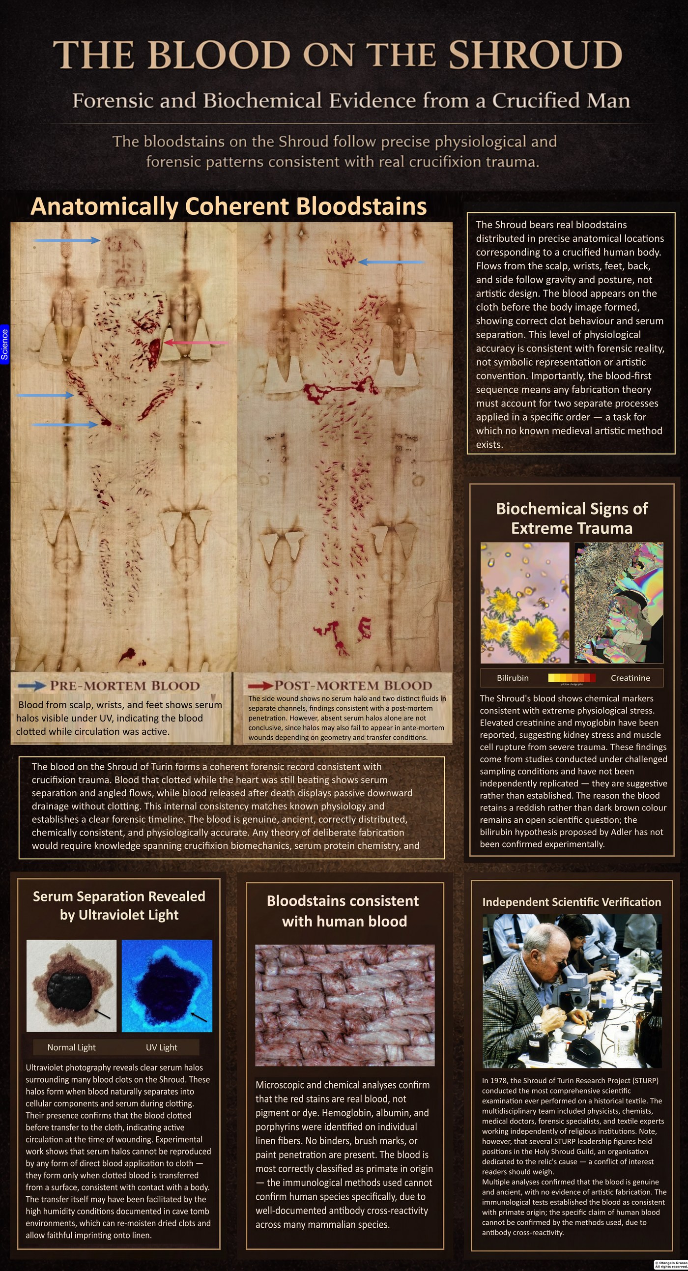
The reddish-brown stains on the Shroud are blood. That identification, made with spectroscopy and immunological tests in the early 1980s, is among the firmest results of the scientific study of the cloth. What the blood can tell us about the death of the man it came from is a harder question.
The stains cover the scalp, the wrists, the forearms, the feet, the side, and the back. Their shapes match the flow of blood from wounds on a body, and in some places the blood is layered in a way that the body’s position would produce. This chapter covers how the stains were identified, the dispute over their origin, the features that have been read as signs of a living or a dying body, and the points on which the evidence remains open.
Identifying the stains
In the late 1970s, during the Shroud of Turin Research Project (STURP) investigation, the chemist John Heller and the physical chemist Alan Adler examined fibres lifted from the stains with adhesive tape. Using a microspectrophotometer, they recorded a strong absorption peak near 410 nanometres. This is the Soret band, the signature of heme, the iron-containing molecule in haemoglobin.1 The peak identified the reddish material as blood rather than a mineral pigment.
Heller and Adler then tested the same material in several independent ways. Immunological assays detected serum albumin and immunoglobulins, proteins that are characteristic of blood.1 Baima Bollone and colleagues reported human haemoglobin and blood-group markers in the stains in 1981.2 Protein-digesting enzymes dissolved the stained fibres, which is what would happen with blood but not with a mineral paint. The analysis also found no binding medium, the oil, egg, or gum that paint needs to stick to a surface. The stains had soaked into the linen rather than sitting on top of it.
The dispute over iron
Not everyone accepted the blood identification. The microscopist Walter McCrone examined tape samples independently and reported iron, which he took to mean a red iron-oxide pigment consistent with a medieval painting.3 His view became the best-known alternative to the blood hypothesis.
The disagreement was about the origin of the iron, not about whether iron was present. Seeing iron is not enough to say what it is bound to. Heller and Adler’s tests showed that the stained material behaved like blood: it had heme chemistry, it reacted with blood-protein antibodies, it dissolved in protein-digesting enzymes, and it had no binding medium. Iron is also present in haemoglobin, so its presence is consistent with blood. Heller and Adler were also aware of iron-oxide particles on the cloth, and they did not deny them. The question was whether those particles were the main constituent of the stains. The tests weighed against that.1
Blood before image
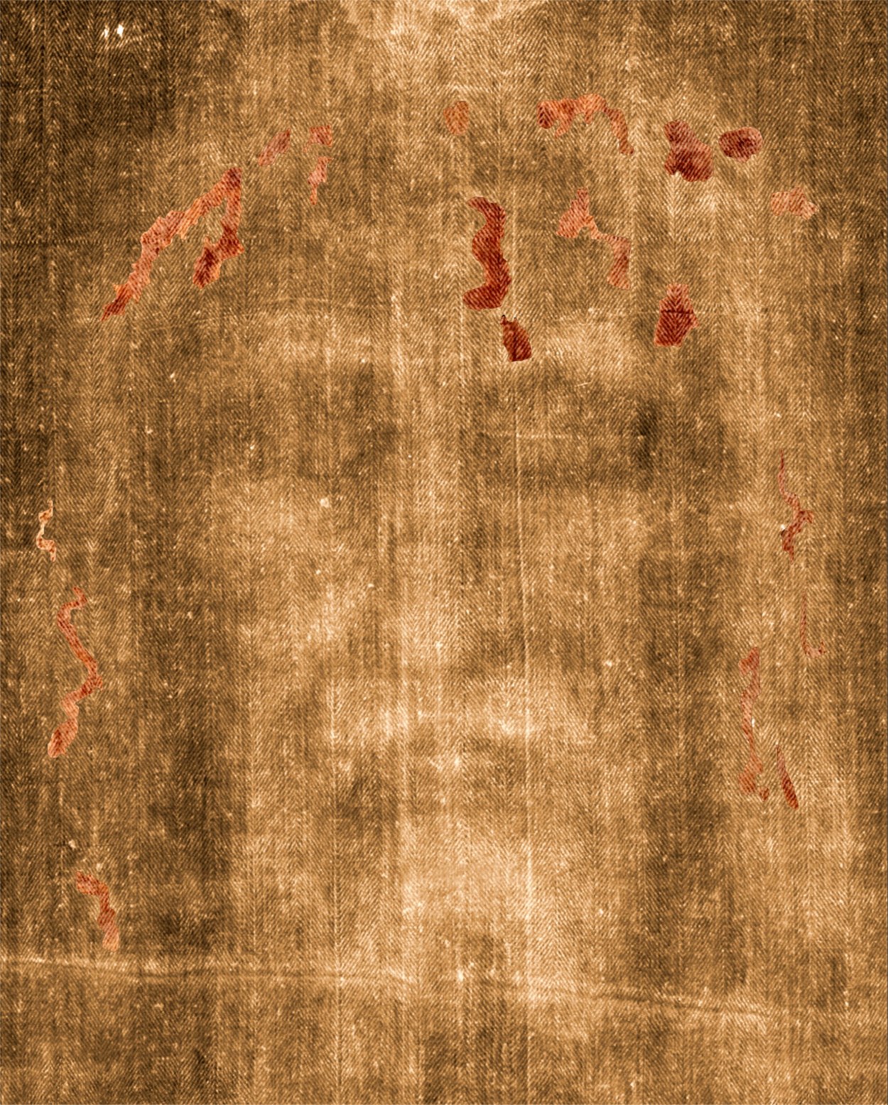
Under magnification, the edges of the bloodstains meet the faint body image in a consistent way. Where blood saturates the linen, the body image stops. The blood was on the cloth before the image formed, and the blood shielded the fibres beneath it from whatever process formed the image.4
This order matters for any account of how the cloth was made. An artist who painted the image first would have had to place the bloodstains with great precision, avoiding every area already marked. An artist who applied the blood first would have had to paint around each clot edge and rivulet. Neither method would be natural for a forger, and the observed order is the one a burial with a body would produce. The order does not identify the process that formed the image, which remains an open question covered in a later chapter.
The colour of the blood
Old blood is normally brown. The Shroud’s stains are red, and skeptics have used that colour as evidence of paint. Adler proposed that the colour could come from bilirubin, a breakdown product of red cells, which accumulates in the blood of people under extreme trauma and may stabilise the red colour over time.
The bilirubin hypothesis has not been confirmed. Experiments that loaded blood with high bilirubin did not reproduce the persistent red colour, and bilirubin is light-sensitive, so its survival for two thousand years is hard to explain. Adler’s proposal involved bilirubin bound to heme derivatives, which may behave differently from free bilirubin, and that version has not been tested directly. Critics have made similar objections on chemical grounds. The colour therefore remains an open chemical question, and it should not be used as an argument for or against authenticity until it is resolved.
Biochemical signs of trauma
Some researchers have reported elevated creatinine, urea, and myoglobin in samples from the Shroud. Creatinine can indicate kidney stress from blood loss, urea can indicate muscle breakdown, and myoglobin can indicate rupture of muscle cells under severe strain. Together these would fit a body in shock.
These results come from sampling that was not done under modern chain-of-custody conditions, and they have not been independently replicated. They are suggestive, not established.
Species, sex, and blood type
Three claims about the blood are often repeated: that it is human, that it comes from a man, and that it is type AB. Each rests on tests that have limits.
Species. The antibodies used in the early studies were labelled “anti-human”, but that label describes how they were made, not how specific they are. Antibodies of this kind can react with blood from other mammals, so the 1981 tests show that the blood was mammalian, and the strongest claim they support is primate origin. The claim of a specifically human source has not been established by the tests conducted.2
Sex. The male result came from PCR tests for Y-chromosome DNA. A beta-globin signal does not show that the DNA came from blood cells, since that gene is also present in skin. A second study found DNA from more than one individual, which points to contamination. Because of these problems, the sex claim is not established either.
Blood type. The AB result is often presented as statistically remarkable, since AB is the rarest common blood type and has been reported for other relics. The statistic is weaker than it looks. Reverse typing relies on antibodies that break down in old samples, which can produce a negative result for any type. The A and B antigens are also carbohydrates found on many bacteria, so a serological result can be produced by microbial contamination. The AB claim needs DNA-based testing before it can be relied upon.
Serum halos and the clock in the cloth
Under ultraviolet light, some stains have been described as showing faint surrounding halos, interpreted as serum-rich areas. That interpretation is not a direct clock: fluorescence alone cannot establish when clotting occurred, whether transfer was direct, or whether the person was alive when the stain formed. Experiments published by König and colleagues reproduced some similar-looking stains using controlled transfers of serum and blood, while noting that such experiments cannot resolve the Shroud’s history.5
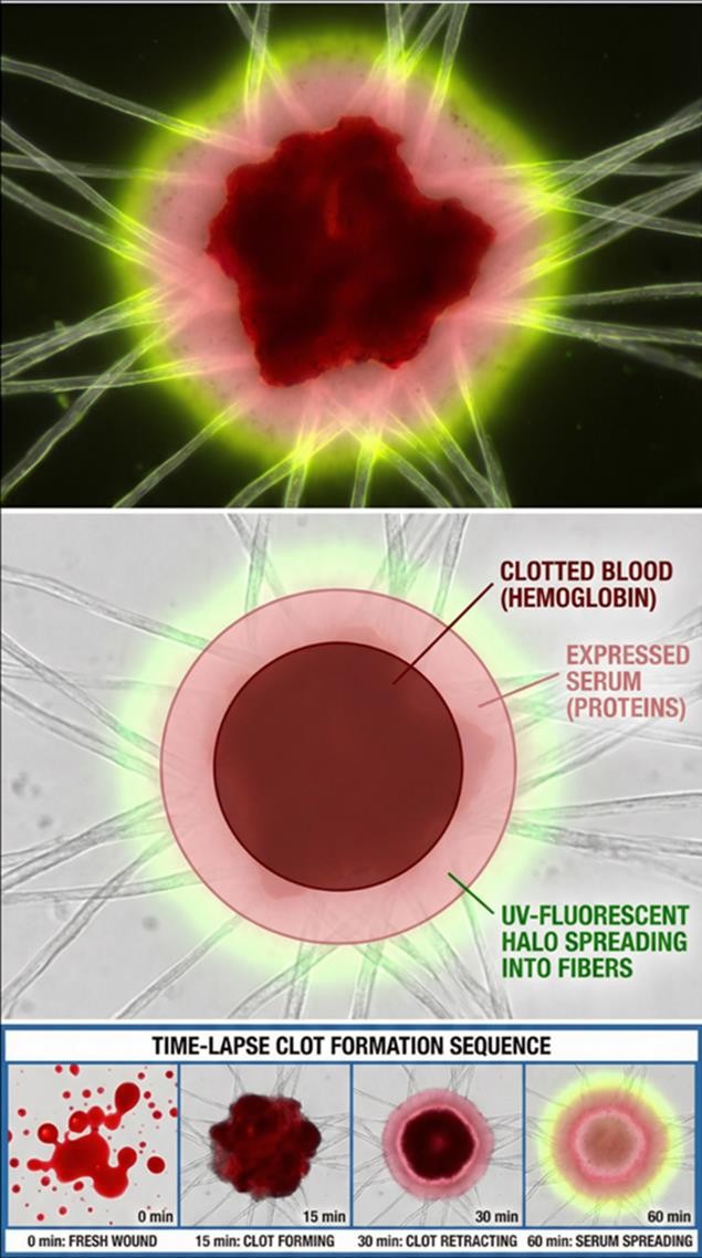
Experimental work has explored how blood and serum behave when transferred to cloth, but resemblance between a laboratory stain and a Shroud stain does not identify a unique transfer process. In particular, halo appearance cannot by itself show that a body was involved in the transfer.
Fluorescence can be affected by the stain’s composition, age, substrate, and illumination. Without a validated comparison under matched conditions, it should not be used alone to infer the age or sequence of a stain.
Blood that beat and blood that pooled
The halo test separates blood that was still circulating when it left the body from blood that seeped out after death. Blood from a living body clots, retracts, and produces serum. Blood from a body that has died is often fluid and does not clot in the same way. Readings of the Shroud’s stains have placed the scalp wounds, which show the strongest halos, in the time when the heart was still pumping strongly. The scourge marks show intermediate halos, and the nail wounds show less. The side wound shows little or none.6
The side wound is the most debated. Its flow pattern and the separation of two fluids have been read as blood that drained after death. Ultraviolet fluorescence around the stain has been interpreted as serum edges. Some of this is interpretation rather than measurement, and the absence of a halo does not prove a wound was made after death, since clotting can fail in a living body and the shape of the body can prevent a halo from forming. The side wound is a candidate for a post-mortem injury, and it is not settled.
Was the body washed?
Some writers have argued that the body was washed before it was wrapped, so that the stains on the cloth reflect seepage after death rather than blood that clotted on the body. Zugibe, a forensic pathologist, argued that the blood visible on the cloth was post-mortem seepage.6
Halo appearance and stain sharpness do not decide between direct contact, transfer from an intermediate surface, or another process. Laboratory studies can test whether a proposed mechanism is physically possible, but they cannot establish that it occurred in this case. The blood evidence should therefore be read as observations with several possible explanations, not as a single verdict.
The paradox that remains
The bloodstains are remarkably sharp. Wicking patterns, serum edges, and the course of the rivulets are all well preserved. A cloth lifted from a body with tacky, half-dried blood would be expected to smear, and the clots would be expected to drag. The sharpness is hard to reconcile with a simple unwrapping.
One proposed mechanism is that dried blood was re-moistened in the humid conditions of a burial cave and transferred to the cloth through capillary action, rather than by direct wet contact. Experiments reported to support this produced smooth, crisp transfers under high humidity and little transfer under low humidity. This may explain some of the sharpness, but not all of it, and it has not been tested on the Shroud itself.
What the blood can and cannot tell us
The blood cannot name the man it came from. Two thousand years of contamination limit what DNA can show about identity, and modern forensic standards would not be met. The blood cannot settle questions of meaning, theology, or the resurrection, which belong to other forms of inquiry.
What the blood can show is more limited, and still significant. It is blood, and its chemistry is consistent with the physiology of severe trauma. It was deposited in an order that matches a body being wrapped in linen. The blood and the image are physically related, and any theory of the cloth has to account for both. Crucifixion physiology, serum chemistry, and post-mortem fluid behaviour are all involved, and a theory of deliberate fabrication would need knowledge of all three before those fields existed in their modern form.
The wounds described in this chapter are read from the stains, and the Passion sequence used here follows the Gospel accounts. The chapters on the scourging, the crown of thorns, and the crucifixion examine each wound in turn.
References
- Heller, J. H., & Adler, A. D. (1981). A chemical investigation of the Shroud of Turin. Canadian Society of Forensic Science Journal, 14(3), 81–103.
- Baima Bollone, P., Jorio, M., & Massaro, A. L. (1981). La dimostrazione della presenza di tracce di sangue umano sulla Sindone. Sindon, 30, 5–8.
- McCrone, W. C. (1990). The Shroud of Turin: Blood or artist’s pigment? Accounts of Chemical Research, 23(3), 77–83.
- Schwalbe, L. A., & Rogers, R. N. (1982). Physics and chemistry of the Shroud of Turin: A summary of the 1978 investigation. Analytica Chimica Acta, 135, 3–49.
- König, T., et al. (2024). Some experiments and remarks regarding the possible formation of blood stains on the Turin Shroud: stains attributed to the crown of thorns, the lance wound and the belt of blood. International Journal of Legal Medicine, 138(1), 229–238. doi:10.1007/s00414-023-02959-6. The study tests possible stain formation; it does not establish the Shroud’s authenticity or a unique transfer process.
- Zugibe, F. T. (2005). The Crucifixion of Jesus: A Forensic Inquiry. New York: M. Evans and Company.